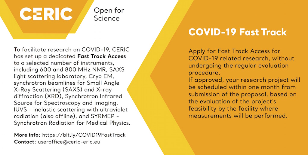
COVID-19 Fast Track Access

In order to facilitate the research on the COVID-19, CERIC has set up a dedicated Fast Track Access to a selected number of instruments.
The dedicated Fast Track Access will allow to access the selected instruments for research related to the COVID-19, without the necessity to undergo the regular evaluation procedure, and be scheduled within one month from the submission of the proposal, based on the feasibility evaluation performed by the facility.
Instruments available:
NMR at the National Institute of Chemistry in Ljubljana:
- ASKA – 600 MHz NMR Spectrometer
- LARA – 600 MHz NMR Spectrometer
- MAGIC – 600 MHz NMR Spectrometer
- DAVID – 800 MHz NMR Spectrometer
TUGraz facility
Synchrotron facility Elettra in Trieste
- X-ray diffraction beamline XRD1
- X-ray diffraction beamline XRD2
- Material characterisation by x-ray diffraction beamline MCX
- Synchrotron Infrared Source for Spectroscopy and Imaging – SISSI BIO
- Offline Synchrotron Infrared Source for Spectroscopy and Imaging – SISSI-BOFF
- Synchrotron Radiation for Medical Physics – SYRMEP
- Inelastic Scattering with Ultraviolet Radiation – IUVS
- Offline inelastic scattering with ultraviolet radiation – IUVS-OFF
SOLARIS National Synchrotron Radiation Centre in Kraków
The Italian Network for Micro and Nano Fabrication:
Contact Person: Luca Ortolani
TEM FEI Tecnai F20
- Facility: transmission and scanning transmission electron microscope operating between 200 and 80kV
equipped with a Schottky Field-Emitter, a Gatan Multiscan CCD camera, a HAADF STEM detector (Fischione), a SUTW EDS detector (EDAX), an electron bi-prism for holography and interferometry and heating and cooling sample holders. - Possible analyses: High Resolution Electron Microscopy (HREM) allowing structural characterizations with a resolution of 0.24 nm, Selected Area Electron Diffraction (SAED) for crystallographic phase determination and electron crystallography, Convergent Beam Electron Diffraction (CBED) for quantitative strain determination, off-axis Electron Holography for compositional and electric fields mapping, Scanning Transmission Electron Microcopy (STEM) coupled with Energy Dispersive X-ray Spectroscopy (EDX) for qualitative and quantitative composition profiles and mapping.
FE-SEM Zeiss 1530
- Facility: scanning electron microscope operating between 0.1 and 30kV, equipped with InLens, Everhart&Thornley SE, BSE and TE detector with scintillator and photo-multiplier, X-Ray spectrometer INCA and two Kleindiek nano-manipulators adapted for in-situ low noise electrical measurements.
- Possible analyses: simultaneous Secondary Electron (SE), Back Scattered Electron (BSE) and Transmitted Electron (TE) analysis. High resolution imaging with in-lens SE detector. EDX analysis and
mapping of composition, electron tomography.
Environmental SEM Zeiss EVO LS10
- Facility: scanning electron microscope operating between 0.1 and 30keV, equipped with secondary and backscattered electrons detectors and X-Ray spectrometer, and capable to operate up to 3000 Pa with non-conductive and wet samples. Cryo stage available.
- Possible analyses: simultaneous SE and BSE imaging on conductive, non-conductive and wet samples. EDX analysis and mapping of composition.
Dual Beam FE-SEM with Focused Ion Beam (FIB) Zeiss XB340
- Facility: equipped with Raith Electron Beam Lithography system, Gas Injection System equipped with Pt, C, XeF2, SiO2 precursor for selective etching and deposition, Kleindik nanomanipulators.
- Possible analyses: SE and BSE investigations coupled with Secondary Ions imaging. High resolution imaging with in-lens SE detector. Slice-and-view 3D reconstruction. Electron and Ion beam Lithography. Ion beam micromachining. TEM lamellas preparation.
Fully equipped TEM sample preparation laboratory
- Facility: mechanical lapping machines, dimpler, variety of metallographic cutting and polishing equipment for sectioning samples, metal and carbon sputtering, equipment for preparing 3 mm discs such as ultrasonic cutter, etc etc) with two ion-milling systems (Gatan PIPS and Gatan DUO-MILL).
Rigaku Smartlab diffractometer
- Facility: multi-purpose 6-axis diffractometer with 9 kW rotating anode Cu Kα source, including the in- plane diffraction attachment and multicrystal monochromators and analyzers optics. The cross beam optics allows rapid switching between parafocusing geometry for powder samples (Bragg-Brentano geometry) and parallel beam geometry for thin films and epilayers. The in-plane diffraction attachment provides an additional detector scanning axis orthogonal to the theta/2-theta diffraction plane to allow measurement of lattice planes perpendicular to the sample surface (e.g., in-plane reciprocal space maps and full pole figures). Two incident beam monochromators {Ge(220) 2-bounce, Ge(220) 4- bounce} and the analyzer monochromator {Ge(220) 2-bounce} enable medium and high resolution XRR and XRD measurements of highly perfect crystals, epilayers, and multilayer samples with up to 12 arc second resolution. X-ray diffraction/reflectivity can be carried out under controlled conditions by means a home-made chamber where nitrogen or hydrated nitrogen can be fluxed inside.
- Possible analyses: (1) determination of structure, stress, texture and phase analysis for a wide range of materials (soft and hard condensed matter) in different forms (bulk, thin film, nanoparticles, and powder), by means of X-ray Diffraction (XRD); (2) investigation of the electron density depth profile of films (crystalline or amorphous) and the surface/interface roughness, by means of X-ray Reflectivity (XRR); (3) detection of the nanoparticle size and morphology and spatial correlation by Grazing Incidence Small Angle X-ray Scattering (GISAXS).
Contact Person: Massimo Cuscunà
FE-SEM Zeiss Merlin
- Facility: Scanning electron microscope operating between 0.1 and 30kV, equipped with InLens, Everhart&Thornley SE. The system is equipped with charge compensation system and insertable annular dark and bright scanning transmission electron microscopy (STEM) detector.
- Possible analyses: simultaneous Secondary Electron (SE) and Transmitted Electron (TE) analysis. High resolution imaging with in-lens. The unique charge compensation system of Merlin also allows the high- resolution imaging of non-conductive samples.
AFM
- Atomic force microscope operating in non-contact and contact mode.
Confocal Microscope
- Confocal microscopy offers several advantages over conventional widefield optical microscopy, including the ability to control depth of field. On an image captured with confocal optics, areas in focus are highlighted. This is called optical sectioning. There is no interference of undesirable scattered light from out-of-focus areas at the highlighted sections. One can therefore get a high-contrast high-resolution image.
Contact Person: Giancarlo Pepponi
D-SiMS
- Secondary ion mass spectrometry (SIMS) is a technique used to analyze the composition of solid surfaces and thin films by sputtering the surface of the specimen with a focused primary ion beam and collecting and analyzing the secondary ions ejected.
The mass/charge ratios of these secondary ions are measured with a mass spectrometer to determine the elemental, isotopic, or molecular composition of the surface to a depth of 1 to 2 nm.
SIMS is presently the most sensitive surface analysis technique, with elemental detection limits ranging from parts per million to parts per billion. The sequential detection of masses simultaneously generated makes the technique best suited for depth-profiling. The most important feature is in fact its ability to follow elemental depth distributions with very low detection limits and with high depth resolution.
The instrument is equipped with low-impact energy – mass filtered – O2 and Cs beams, a magnetic sector mass spectrometer (energy filtered), electron beam for charge compensation, rotating sample holder and laser interferometer for continuous monitoring of the sputtering rate.
The overall design of this instrument minimizes ion beam induced effects (such as mixing and roughness development) during depth profiling, allowing to reach depth resolutions as good as 0.8 nm/decade.
• Ion sources: Oxygen and Cesium
• Impact energy: 0.2÷15 keV
• Analyzer: Magnetic Sector
• Min depth resolution: 1.5 nm, 1÷20 nm typical
• Mass resolution δM/M: up to 20.000
• Mass range: > 500 amu at 5 kV sec. ext. Volt.
• Spatial resolution: 1 μm
• Charge compensation: provided by electron flooding
• Det. limit (deep profile): B, P and As < 1015 at/cm3
• Det. limit (shallow profile): B, P and As < 1016 at/cm3
• Scan limits: 500 μm x 500 μm
ToF SiMS
- Time of Flight Mass analysers are typically combined with a “static” approach to Secondary-Ion Mass Spectrometry. Static SIMS is a technique for chemical analysis including elemental composition and chemical structure of the uppermost atomic or molecular layer of a solid. ToF-SIMS provides 2/3-dimensional spatial distributions of both elements and chemical compounds (through the analysis of ejected molecular ions) on virtually each kind of solid materials, i.e. with no limitations with respect to insulating, organic or easily damaged samples.
The term ‘static’ is used to indicate that in this case the ionic fragments come from an unchanged surface (practically unaffected by implantation and mixing typically present in ion bombardment). A wide range of applications of the technique are therefore found in the analysis of polymers (the packaging industry is perhaps the leading example in this respect), organic materials, biological samples, that is all those areas which are not or only hardly accessible to the traditional mass spectrometric techniques.
• Ion sources: Bismuth
• Impact energy: 25 keV
• Ion sputtering: Argon, Cesium, Oxygen, Xenon (1-10 keV)
• Mass resolution δM/M: 8.000
• Mass range: typically 10.000 (no limit)
• Spatial resolution: 80 nm
• Charge compensation: provided by electron flooding
• Heatable/coolable stage
• Analyser: Reflectron parallel detection
• Depth Resolution: 1-3 monolayers (static mode)
• Detection Limit: B, P and As < 1015 at/cm3
PTR-MS
-
- PTR-MS is the abbreviation for Proton Transfer Reaction – Mass Spectrometry. The technology is based on reactions of ion donors (usually H3O+), which perform non-dissociative proton transfer to all Volatile Organic Compounds (VOCs) with higher proton affinity. These characteristics result in sensitivity allowing the detection of VOCs in air in the pptv range. The very soft ionization technique avoids mass fragmentation, which enhances the interpretability of mass spectra. A TOF-MS linked to the PTR system offers real-time, high time resolution, high sensitivity detection of all analytes in parallel. There is no need for time consuming sample preparations or chromatographic separation before injection into the PTR MS inlet.
• Source: Proton Transfer Reaction/Atmospheric Pressure Chemical Ionisation
• Analyser: Time of Flight Reflectron for parallel detection • Mass resolution δM/M: 1300
• Compounds detectable: All molecules with proton affinity > H2O
• Detection limit: few ppt
XPS
- X-ray photoelectron spectroscopy (XPS) also known as ESCA (Electron Spectroscopy for Chemical Analysis) is a surface-sensitive quantitative spectroscopic technique that measures the elemental composition at the parts per thousand range.
Not only elemental analysis is provided but also chemical state and electronic state of the elements in the near surface region of the material.
XPS spectra are obtained by irradiating a material with a beam of X-rays while simultaneously measuring the kinetic energy and number of electrons that escape from the uppermost layer (~10 nm) of the material being analyzed.
XPS is a surface chemical analysis technique that can be used to analyze the surface chemistry of a material. In principle XPS detects all elements. In practice detects all elements with an atomic number (Z) above 3 (lithium).
XPS is routinely used to analyze inorganic compounds, metal alloys, semiconductors, polymers, catalysts, glasses, ceramics, paints, papers, inks, woods, plant parts, make-up, teeth, bones, medical implants, bio- materials, viscous oils, glues, ion-modified materials and many others.
• X-Ray Source: Monochromatic AlKa radiation
• Analyzer: Hemispherical Analyzer,
• Transmission or Spatial mode
• Sample imaging: via optical microscope
• Energy resolution δE: < 0.3eV on the Ag Fermi edge with a pass energy of 75 eV
• Sensibility: 0.5 – 1 at%
• Spatial resolution: 10μm. XPS line scans/maps can be acquired.
• In-depth information Ar gun (up to 5 keV) for etching process.
• Wet etching to preserve chemical information.
• Charge compensation: provided by a low energy electron flooding.
AFM
The AFM is member of the family of Scanning Probe Microscopes (SPMs). The sensing element of those microscopes is a sharp tip mounted on a cantilever and slide on the surface of the specimen. The interaction forces between the tip and atoms of the sample surface (approximately nano-newton) cause a deflection of the lever on which the tip is mounted. Following a change of topography, a change in the deflection of the lever happens. A laser that hits the back side of the cantilever, is reflected towards a 4 quadrant photodiode. Detailed topography maps of the surface are obtained by moving the probe by means of piezoelectric actuator. Several scanning modes are usually available, whose difference lays in the type of movement (e.g. continuous or oscillating), the distance at which the scanning is done (e.g. in contact with the surface or at a certain distance from it), and the type of signal recorded (e.g. normal or lateral deflection).
The great advantage of the AFM technique is that analyses are performed in air and, unlike STM, it allows microscopy on insulator materials. Besides it is a non-destructive analysis and it does not demand particular preparations of the samples.
SOLVER P47H AND SOLVER Px
Instruments in FBK are equipped with high resolution head and electric head that allows EFM (Electrostatic force microscope) and SKM (Scanning Kelvin Microscopy) measurements also known as surface potential microscopy. It is also equipped with controlled gas environment.
• Measuring modes:
o Contact AFM/ LFM/ Resonant Mode o Semicontact + Noncontact AFM
o Phase Imaging
o Force Modulation (Viscoelasticity)
o MFM/ EFM/ SKM
o AFM Lithography-Force
• High Res Scan (X-Y-Z): 50 x 50 x 4 μm
• High Res Vertical Resolution: < 0.5 Å
• Electrical Meas Scan (X-Y-Z) : 90 x 90 x 5 μm
• Max. Sample Size: 15mm Ø
Contact Person: Riccardo Bertacco
AFM System – Keysight 5600LS
- Imaging Modes: The 5600LS is compatible with contact mode, acoustic AC mode, phase imaging, STM, LFM, KFM/EFM, MFM, force modulation, current sensing, and MAC Mode, a nondestructive technique for imaging delicate samples in air or liquid.
The machine is compatible with the investigation of biological samples in a Petri dish.
XPS-UPS System – Phoibos 150 hemispherical analyzer
- Photon sources: XPS: 1253.6 eV (Mg-Ka) and 1486.6 eV (Al-Ka) UPS: 21.2 eV (He-I) and 40.8 eV (He-II) Sample size: 1×1 cm max
Contact person: Sara Pascale
Availability to analyze safety group 1 samples (agents that do not pose a risks of causing disease in human beings) with the following instrumentation:Optical Microscope: Nikon Eclipse L200N
- Magnification: 50X – 1500X
Dark Field, Bright Field, phase contrast prism Filters: ND4, polarizer
Fluorescence Illumination C-HGFI HG Fiber Illuminator “Intensilight” MBF72655 Automatic acquisition of large images in .nd2 format (NIS-Elements D 4.13.04)
Scanning Electron microscope (SEM): Tescan MIRA 3 FEG-SEM
- Scanning electron microscope operating between 0.1 and 30 kV. Samples must be vacuum-compatible.
Imaging modes: low vacuum mode, low voltage imaging. - Available detectors: ▪ Secondary election detector (SE, external and in-beam)
▪ Backscattered electron detector (BSE, annular and in-beam detector)
FilmTek 4000 NIR
The system measures the optical material properties of the thin films:
- Analysis of refractive index in the 380 nm – 1700 nm wavelength range, large area mapping possible.
- Multi-Angle Differential Power Spectral Density algorithm enabling measurement of index with a resolution as high as 0.00002, independent measurement of thickness and index, and independent measurement of TE and TM components of index.
- Analysis of planar samples min. 2×4 mm in size.
Profilometer Tencor KLA P-7
- The instrument can characterize morphologically hard or soft samples with 2D and 3D mapping modes. The measurable differences in height are between 0.01 and 1000 μm. The mapping is carried out with contact-mode height profiling. The sample must be able to withstand a pressure of 0.03 mg minimum applied to the scanning tip with a radius of approx. 20 μm.
Please note that since these facilities do not have a special bio-safety accreditation, each project applicant is expected to consider this fully when submitting a proposal and in the later stage samples, and to disclose all data relevant for safety of the staff at individual facility. Only samples guaranteed as non-harmful and with no ability to cause or transfer viral infection can be accepted for research.
Due to the travel restrictions in place for several countries, remote access will be preferred.
Proposals have to be submitted through the VUO. After registration/login, follow the dedicated link “Submit a new FAST TRACK related to COVID-19 research”.
For more information, also regarding the commercial use, please contact useroffice@ceric-eric.eu
As a pilot of open access of the H2020 project ACCELERATE, all scientific information generated must be available and reusable through online access that is free of charge to the end-user. ‘Scientific information’ can mean:
1. peer-reviewed scientific research articles (published in scholarly journals), or
2. research data (data underlying publications, curated data and/or raw data).
‘Access’ includes not only basic elements – the right to read, download and print – but also the right to copy, distribute, search, link, crawl and mine.
The 2 main routes to open access are:
Self-archiving / ‘green’ open access – the author, or a representative, archives (deposits) the published article or the final peer-reviewed manuscript in an online repository before, at the same time as, or after publication. Some publishers request that open access be granted only after an embargo period has elapsed.
Open access publishing / ‘gold’ open access – an article is immediately published in open access mode. In this model, the payment of publication costs is shifted away from subscribing readers.
-
24.07.2024
21st CERIC Call for Proposals now OPEN
-
16.07.2024
Read the CERIC newsletter | July 2024


