

Materials Characterisation by X-ray Diffraction (MCX)
Elettra Sincrotrone Trieste
X-ray diffraction is a tool used for identifying the atomic and molecular structure of a crystal, in which the crystalline atoms cause a beam of incident X-rays to diffract into many specific directions. By measuring the angles and intensities of these diffracted beams, a crystallographer can produce a three-dimensional picture of the density of electrons within the crystal. From this electron density, the mean positions of the atoms in the crystal can be determined, as well as their chemical bonds, their disorder and various other information of different materials, especially minerals and alloys. The MCX beamline can perform a wide range of non-single crystal diffraction experiments, from grazing angle diffraction and reflectivity through residual stress and texture analysis to phase identification, structural studies and kinetic studies. Systems that can be investigated vary from organic and inorganic thin films through thermally and/or mechanically modified surfaces of mechanical components to polymers or catalysts. Highly disordered materials in the form of films, powders and fibers can also be investigated.
Contact: Jasper Plaisier
Tel: +39 040 375 8714 (office) | +39 040 375 8751 (beamline)
Technical specifications
The double crystal monochromator (DCM) consists of two Si crystals (active area 50×50 mm2, manufactured cut along the [111] direction, which can be precisely positioned and oriented in the X-ray beam. Two successive Bragg reflections with an inherent energy resolution of 0.014% (given by the Darwin angular width of the Si 111 reflection) will direct photons of the desired energy parallel to the incoming beam direction, but offset upward (out of the direct Bremsstrahlung beam). This “fixed exit” operation is achieved by placing the crystals on two independent rotation stages, and translating crystal 2 along the beam direction. The second crystal provides sagittal focusing; it is a ribbed crystal, cylindrically bent to a variable curvature radius.
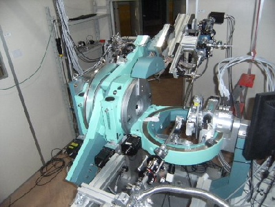
Sample environment
The experimental setup consists of a Huber goniometer with k geometry and is fully controllable from remote. The beamline hosts small molecules, protein crystallography, powder diffraction, high pressure physics and solid-state experiments.
Detailed information can be found on the beamline’s main homepage.