

Surface graphitization by microfocused soft X-rays and electron beams
|Since the beginning of the XX century, our data storage is based on magnetic devices, from magnetic tapes to hard disks. In these devices, the information is saved as a magnetized sector of the tape, or disk, and the technological advancements allowed their miniaturization. As an example, the areal density increased of five hundred million times from 1956 to 2014, having an impact also on the cost per gigabyte, which decreased from nine million dollars to two cents from 1957 to 2018, thus allowing for the construction of small but powerful devices, such as smartphones and tablets. To achieve further advancements is fundamental to be able to manipulate magnetic regions with new techniques.
CERIC project MAG-ALCHEMI aims at developing tools to control thin-film magnetism via interfacial engineering. The capability to locally control the magnetic state is pursued trough lithographic methods. In fact, lithography allows to sculpt structures with arbitrary shapes by both removing or adding atomic species on surfaces with remarkable accuracy.
A research by Francesca Genunzio, Andrea Locatelli, scientists at the CERIC synchrotron facility in Trieste (ITA), and colleagues, highlighted the possibility to use photons and electrons to manipulate a magnetic substrate at the nanometric scale. Researchers were able to deposit a layer of carbon atoms over a thin cobalt film. This result was achieved by adsorption of carbon monoxide (CO) followed by microfocused soft X-rays or electron beams. This procedure stimulates the CO dissociation and the release of oxygen, thus leaving a layer of carbon atoms. The process was monitored in real-time by Fast-XPS and XPEEM.
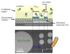
BOTTOM: Low energy electron microscope (LEEM) image showing experimental results
The results show that, in the case of soft X-rays, the dissociation probability increases with the energy of the photons. At the same time, low energy electrons in the range of 50-200 eV, dissociated CO more efficiently than X-rays. After the stimulated CO dissociation, the thin layer was heated, causing the desorption of the carbon monoxide molecules in the nonirradiated regions. The remaining graphitic overlayer modified the magnetic anisotropy of the cobalt film, and protected it from oxidation in ambient conditions, therefore enabling the sample transfer without protection. A remarkable feature for ultra-thin magnetic films.