

Soft X-ray Transmission and Emission Microscope (TwinMic)
Elettra Sincrotrone Trieste
An X-ray microscope uses electromagnetic radiation in the soft or hard X-ray band to produce magnified images of objects. The shorter wavelengths of X-rays allow higher optical resolution than with visible light microscopy. High X-ray penetration power gives ‘deeper’ insight into the specimen and in many cases avoids slicing, as required for electron microscopies. The electronic structure of atoms provides each element with a specific fingerprint that allows X-ray microscopy to identify the elemental distribution. X-ray analytical techniques are even sensitive to slight modification in the electronic structure of an atom by its neighboring atoms, which can provide an additional wealth of information on the chemical speciation of the specimen. TwinMic offers transmission soft X-ray microscopy with an optical resolution up to 10 times higher than conventional visible microscopy, combined with a natural contrast between organic matter and water that allows imaging of specimens in their natural liquid environment without staining. The application of TwinMic ranges over a wide variety of fields, from nanotechnology and biotechnology through environmental science and cultural heritage to clinical and medical applications.
Contact: Alessandra Gianoncelli
Tel: +39 040 375 8753 (office) | +39 040 375 8478 (beamline)
Technical specifications
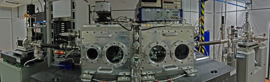
The TwinMic X-ray spectromicroscope is a world-wide unique instrument that combines full-field imaging with a scanning X-ray microscope in a single instrument. The instrument is equipped with versatile contrast modes, including absorption or brightfield imaging, differential phase and interference contrast or Zernike phase contrast – as is customary from a visible light microscope. The microscope is operated in the 400 – 2200 eV photon energy range or as equivalent to 0.56 – 3 nm wavelengths. Depending on the energy and X-ray optics, TwinMic can reach sub-100nm spatial resolution.
Detailed information can be found on the beamline’s main homepage.