

Synchrotron radiation shows a rare disease in a new light*
|Alkaptonuria (AKU) is a rare disease associated with the lack of a specific enzyme with the complicated name homogenisate 1,2-dioxygenase. One of the purposes of this enzyme is to metabolise, meaning chemically transform, Homogenastic Acid (HGA) which occurs when the human body metabolises the essential amino acid tyrosine. If the HGA is not metabolised in the correct way, it develops to Alkapton (1,4-benzoquinon-2-acteic acid). Over the years, HGA and Alkapton form a pigment called “ochronic pigment”, with a chemical structure similar to the dark human pigment melanin. Unlike melanin, it produces oxidising substances that accumulates in collagen rich tissues (e.g. cartilage in ears and knees) and body fluids, leading to chronical inflammation of the interested tissues. In the initial state of the disease, the inflammation only causes a darkening of the cartilage, but in further stages it leads to tissue degeneration and a severe and sometimes crippling form of arthropathy. Although the clinical features of AKU are very well described, little is known about the molecular interactions of the ochronic pigment with the collagen fibres.
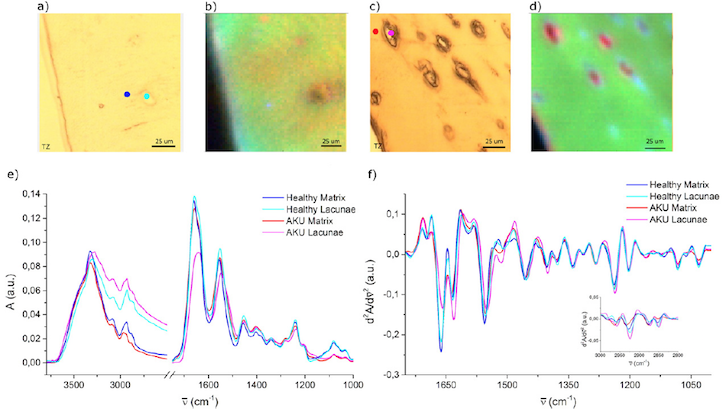
To shed some light onto these interactions, a research team around Elisa Mitri, Lisa Vaccari, Alessandra Gianoncelli and Annalisa Santucci from Elettra Sincrotrone Trieste and the University of Siena investigated AKU cartilage tissue combining synchrotron based Fourier-Transform Infrared Microscopy (FTIRM) and Low Energy X-ray Fluorescence (LEXRF) microscopy at the Italian CERIC Partner Facility in Trieste. The combination of both techniques allows gathering information about the biochemical structure and elemental composition of tissue in a very high resolution at the cellular level. The aim of the study was to compare healthy with sick tissues, to find distinctive biomolecular signatures and elemental compositions for AKU.
During the study, some distinctive features of AKU cartilage have been identified. For the first time it was observed that the ochronic pigment is agglomerated with different kinds of clotted, dysfunctional proteins in the tissues. Furthermore, AKU tissues showed a high amount of sodium and low amount of magnesium, when compared to the healthy tissues. Although the study could not give explanations on why those features occur, the data provide a more detailed picture of the complex and unique pathways of this rare disease. Now it’s the time to study these single pathways, using the same multi technique approach in simpler cellular model systems to solve the puzzle of AKU piece by piece, and design proper therapeutic approaches