

Bioengineered scaffolds to preserve fertility after ovarian tissue transplantation
|In recent decades, breakthroughs in cancer treatments have improved survival rates for young female patients. However, the gonadotoxic effects of chemotherapy and radiotherapy often leave survivors facing premature menopause and infertility, heavily affecting long-term quality of life. For prepubertal girls, ovarian tissue cryopreservation and transplantation represents the first-choice approach. Yet, revascularization remains a challenge, since ovarian grafts experience a critical ischemic window—a period of 5 to 10 days before new blood vessels form and perfuse the tissue.
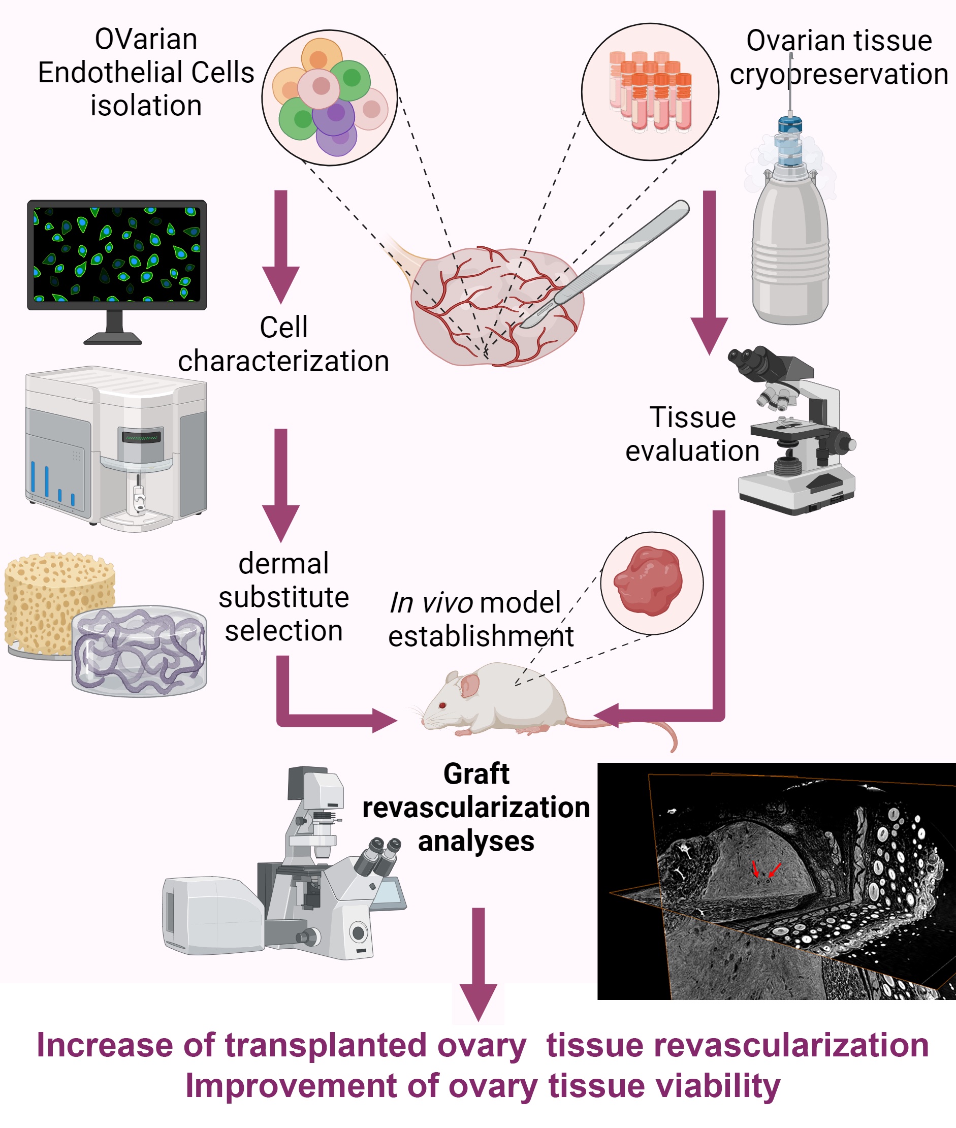
To overcome this problem Mariagiulia Spazzapan (University of Trieste), Chiara Agostinis, Lorella Pascolo (IRCCS Burlo Garofolo) and colleagues tested the pro-angiogenic potential of four different 3D biomaterial scaffolds (engineered dermal matrices that mimic the natural extracellular environment and encourage tissue regeneration), assessing their ability to interact with ovarian endothelial cells (OVEC). The most suitable skin substitute were employed for seeding OVECs and testing transplantation of human ovarian tissue in the animal model.
To investigate the revascularization process researchers used, among other techniques, synchrotron radiation-based phase-contrast microtomography (microCT) at the SYRMEP beamline, available at CERIC Italian Partner facility in Elettra Sincrotrone Trieste. This analytical technique offered non-invasive, high-resolution 3D images that revealed formation of new vascular structures within the scaffold matrix, accumulation of red blood cells in vessel-like formations, clear evidence of OVEC migration into the ovarian tissue, and preservation of healthy primordial follicles post-transplantation. Notably, grafts without endothelial supplementation did not show these vascular features, emphasizing the effectiveness of the OVEC-enhanced scaffold.
These findings mark a significant step forward in fertility preservation for cancer survivors, by combining cryopreserved tissue with endothelial cell-supplemented 3D scaffolds. Moreover, this approach exemplifies the potential of tailored, bioengineered regenerative therapies.
ORIGINAL ARTICLE: