

Particle-Inducted X-ray Emission and Rutherford Backscattering (PIXE/RBS)
Ruđer Bošković Institute in Zagreb
Particle-induced X-ray emission or proton-induced X-ray emission (PIXE) is a technique used in the determination of the elemental make-up of a material or sample. When charged particles (e.g. protons) move through a material, they lose energy primarily by exciting electrons in the material’s atoms. Electrons in the inner shells of the atom (predominantly the K and L shells) gain enough energy to eject. Electrons from outer shells fill these vacancies, which is accompanied by the emission of X-rays. The energies of these X-rays are characteristic of the element and can be used to identify the elemental composition of the sample. PIXE is a relatively simple and multi-elemental analytical technique that can be used to identify and quantify elements ranging from Na to U. Due to the high signal to background ratio, PIXE is also a nondestructive technique and very sensitive, and for a wide range of measured elements, with detection limits close to 1 ppm.
Protons can also interact with the nucleus of the atoms in the sample through elastic collisions, Rutherford backscattering, often repelling the proton at angles close to 180 degrees. By measuring the energy and intensity of the backscattered beam of high energy protons, it is possible to determine the composition and depth profile of elements on the sample surface and below. The technique is in particular powerful for depth profiling of heavy elements in light substrates. When RBS is done in combination with PIXE, it can be used to determine light element concentrations, which is not possible by PIXE alone.
Technical specifications
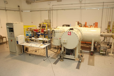
The scattering chamber for routine PIXE/RBS analysis is installed at a -45 degree line of the Tandteron accelerator. It is equipped with two PIXE detectors. The SDD detector is used for the analysis of light elements (Na and above). The Si(Li) detector has a large solid angle and a Mylar filter is optimized for heavy elements (K, Ca and above). Quantification is based on the GUPIX software package, using integrated current or RBS normalization.
Contact: Zdravko Siketic
Sample environment
The chamber has an integrated sample holder, which can take up to 16 samples (size between 10 mm and 25 mm). The samples are exposed to a 2 MeV proton beam of circular shape (3, 5 or 8 mm. Detailed information can be found on the instrument’s webpage.
-
25.08.2025
Microfabrication Laboratory