



Synchrotron light reveals nanoplastics inside human reproductive cells
|Nanoplastics are increasingly being detected in human tissues, from blood and placenta to breast milk and ovarian follicular fluid. However, one key question remains: how can we reliably detect particles that are hundreds of times smaller than a cell once they are inside complex biological tissues?
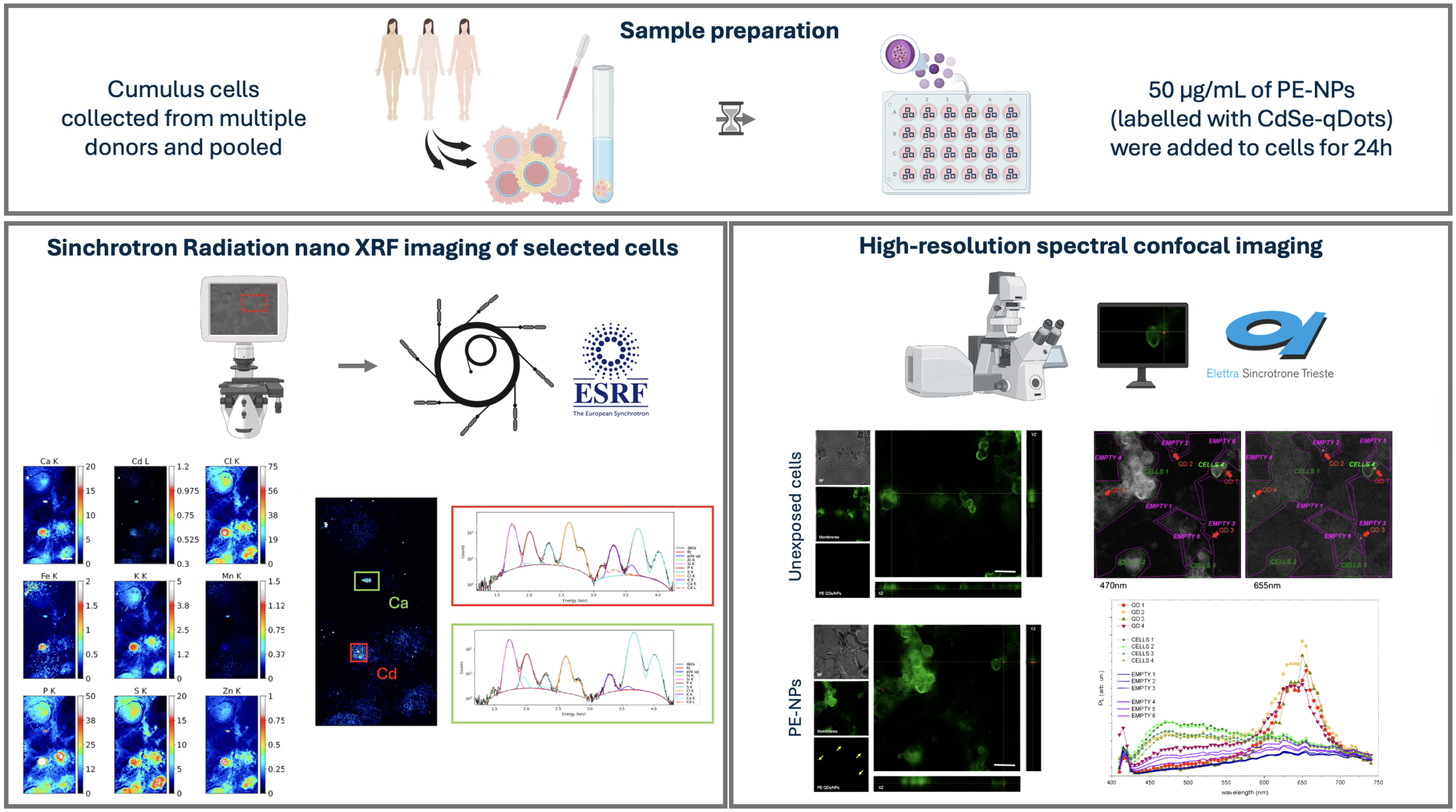
Sara Bozzer, Cristina Tufoni, Lorella Pascolo and colleagues of the Institute for Maternal and Child Health (IRCCS Burlo Garofolo, Trieste – Italy) tackled this challenge by developing an innovative imaging workflow capable of locating polyethylene nanoplastics within human cumulus cells, the specialized cells that surround and support the oocyte. These cells are of particular interest because they represent the first biological interface between environmental contaminants and the female germ cell.
The researchers combined spectral confocal microscopy performed at the CERIC Italian Partner Facility at Elettra with synchrotron-based nanoscale X-ray fluorescence imaging (SR-nXRF) carried out at the ID21 beamline of the European Synchrotron Radiation Facility (ESRF). This complementary approach overcame the limitations of conventional optical imaging and enabled the researchers to detect rare nanoplastic-associated signals and precisely locate cadmium-labelled polyethylene nanoplastics inside intact cells.
This work establishes a powerful framework for future investigations into how nanoplastics interact with human cells: by making the invisible visible, advanced and complementary synchrotron imaging techniques open new opportunities to understand the potential impact of these emerging contaminants on reproductive health and early development.
ORIGINAL ARTICLE: